Ct Head Template
Ct Head Template - Use these templates to create solid preliminary reports and make your personal brand known. 0.625 mm thick axial images as well as coronal and sagittal reformatted. Free download pdf word format ct brain plain. Simplify and enhance your radiology reporting today! Sometimes during the exam you may. Also available other updated radiology mri, ct scan, xray, sonography, usg, mammography, pet ct, eeg and ecg report templates. Explore our growing collection of free radiology report templates in.rtf format, compatible with powerscribe 360. A ct head standard report may not be applicable in all situations, but gives an idea of some of the areas to cover when reporting a ct head. Ct scanning—sometimes called cat scanning—is a noninvasive, painless medical test that helps physicians diagnose and treat medical conditions. Minor small vessel ischaemic disease of the deep white matter is noted. Also available other updated radiology mri, ct scan, xray, sonography, usg, mammography, pet ct, eeg and ecg report templates. Each template has one or more options: 2.5 mm thick axial images were obtained through the head without (or before and after) the use of intravenous contrast. No abnormality of the visualized intracranial compartment or orbits. A ct head standard report may not be applicable in all situations, but gives an idea of some of the areas to cover when reporting a ct head. Many templates incorporate rsna’s radlex® terminology and radelement common data elements developed by rsna, in collaboration with the acr and radiologic subspecialty. Radreport templates are intended to provide examples of best practices for diagnostic reporting. 0.625 mm thick axial images as well as coronal and sagittal reformatted. _______ no intra or extra. Ct scanning—sometimes called cat scanning—is a noninvasive, painless medical test that helps physicians diagnose and treat medical conditions. _______ no intra or extra. We've added it to your search pattern for head ct. Structured radiology report template for a ct of the neck with intravenous contrast. 2.5 mm thick axial images were obtained through the head without (or before and after) the use of intravenous contrast. Free download pdf word format ct brain plain. Free download pdf word format ct brain plain. Simplify and enhance your radiology reporting today! Radreport templates are intended to provide examples of best practices for diagnostic reporting. Many templates incorporate rsna’s radlex® terminology and radelement common data elements developed by rsna, in collaboration with the acr and radiologic subspecialty. No abnormality of the visualized intracranial compartment or orbits. Ct scanning—sometimes called cat scanning—is a noninvasive, painless medical test that helps physicians diagnose and treat medical conditions. Radreport templates are intended to provide examples of best practices for diagnostic reporting. Radreport templates are intended to provide examples of best practices for diagnostic reporting. 2.5 mm thick axial images were obtained through the head without (or before and after) the. Free download pdf word format ct brain plain. Also available other updated radiology mri, ct scan, xray, sonography, usg, mammography, pet ct, eeg and ecg report templates. Also available other updated radiology mri, ct scan, xray, sonography, usg, mammography, pet ct, eeg and ecg. 0.625 mm thick axial images as well as coronal and sagittal reformatted. Brain and bone windows. 2.5 mm thick axial images were obtained through the head without (or before and after) the use of intravenous contrast. Each template has one or more options: Simplify and enhance your radiology reporting today! Sometimes during the exam you may. Free download pdf word format ct brain plain & contrast. 0.625 mm thick axial images as well as coronal and sagittal reformatted. A ct head standard report may not be applicable in all situations, but gives an idea of some of the areas to cover when reporting a ct head. Sometimes during the exam you may. Also available other updated radiology mri, ct scan, xray, sonography, usg, mammography, pet ct,. A ct head standard report may not be applicable in all situations, but gives an idea of some of the areas to cover when reporting a ct head. Structured radiology report template for a ct of the neck with intravenous contrast. Explore our growing collection of free radiology report templates in.rtf format, compatible with powerscribe 360. Ct scanning—sometimes called cat. _______ no intra or extra. Structured radiology report template for a ct of the neck with intravenous contrast. Simplify and enhance your radiology reporting today! Free download pdf word format ct brain plain. 2.5 mm thick axial images were obtained through the head without (or before and after) the use of intravenous contrast. Many templates incorporate rsna’s radlex® terminology and radelement common data elements developed by rsna, in collaboration with the acr and radiologic subspecialty. Radreport templates are intended to provide examples of best practices for diagnostic reporting. Minor small vessel ischaemic disease of the deep white matter is noted. Radreport templates are intended to provide examples of best practices for diagnostic reporting.. Free download pdf word format ct brain plain. Radreport templates are intended to provide examples of best practices for diagnostic reporting. Each template has one or more options: Free download pdf word format ct brain plain & contrast. Minor small vessel ischaemic disease of the deep white matter is noted. 2.5 mm thick axial images were obtained through the head without (or before and after) the use of intravenous contrast. Ct scanning—sometimes called cat scanning—is a noninvasive, painless medical test that helps physicians diagnose and treat medical conditions. Free download pdf word format ct brain plain. Use these templates to create solid preliminary reports and make your personal brand known. We've added it to your search pattern for head ct. Minor small vessel ischaemic disease of the deep white matter is noted. No abnormality of the visualized intracranial compartment or orbits. Many templates incorporate rsna’s radlex® terminology and radelement common data elements developed by rsna, in collaboration with the acr and radiologic subspecialty. Users of radreport are welcome to download the published templates. Each template has one or more options: Explore our growing collection of free radiology report templates in.rtf format, compatible with powerscribe 360. A ct head standard report may not be applicable in all situations, but gives an idea of some of the areas to cover when reporting a ct head. _______ no intra or extra. Free download pdf word format ct brain plain & contrast. Also available other updated radiology mri, ct scan, xray, sonography, usg, mammography, pet ct, eeg and ecg. Brain and bone windows were reconstructed.
Figure 7 from A Neonatal Bimodal MRCT Head Template Semantic Scholar
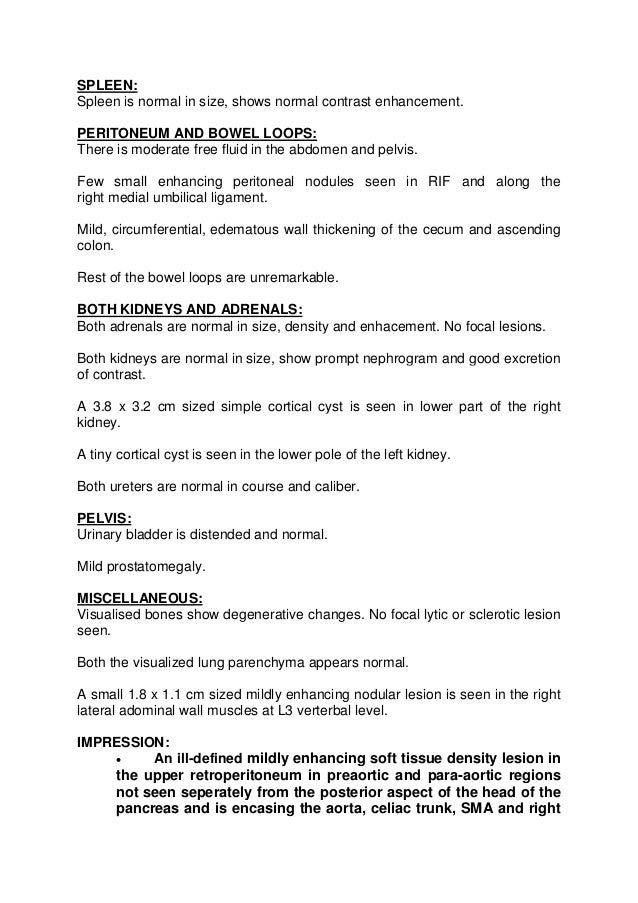
Statim healthcare CT Scan Sample Reports

Approach to CT head
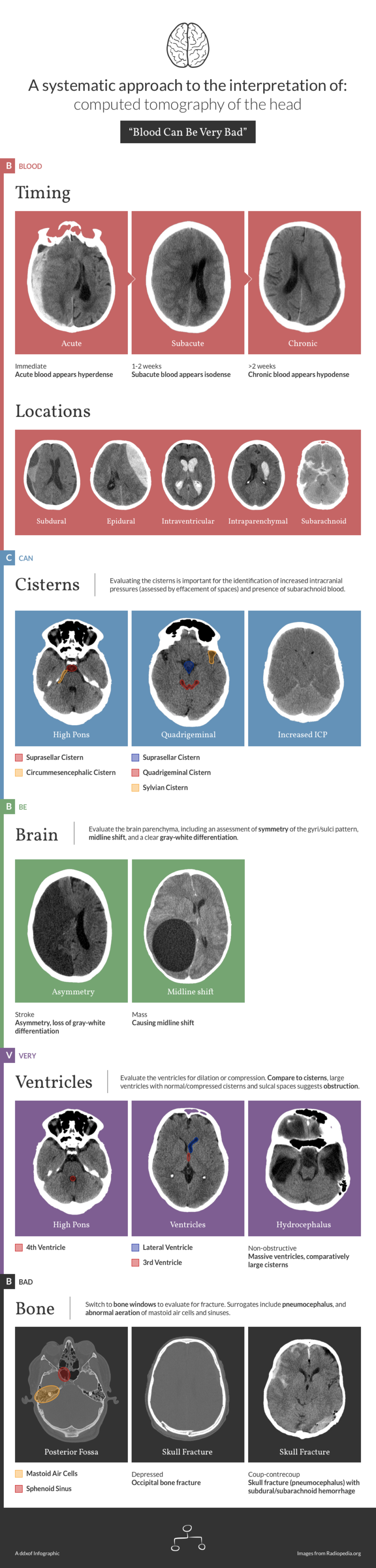
Differential Diagnosis of Infographic CT Head Interpretation

Figure 1 from A Neonatal Bimodal MRCT Head Template Semantic Scholar

Figure 3 from A Neonatal Bimodal MRCT Head Template Semantic Scholar

(PDF) A neonatal bimodal MRCT head template

Figure 2 from A Neonatal Bimodal MRCT Head Template Semantic Scholar

Figure 8 from A Neonatal Bimodal MRCT Head Template Semantic Scholar
![cthead [Open Med School]](https://openmed.school/_media/ct_head_with_contrast_unremarkable.png)
cthead [Open Med School]
0.625 Mm Thick Axial Images As Well As Coronal And Sagittal Reformatted.
Users Of Radreport Are Welcome To Download The Published Templates And To Create.
Sometimes During The Exam You May.
Also Available Other Updated Radiology Mri, Ct Scan, Xray, Sonography, Usg, Mammography, Pet Ct, Eeg And Ecg Report Templates.
Related Post: